

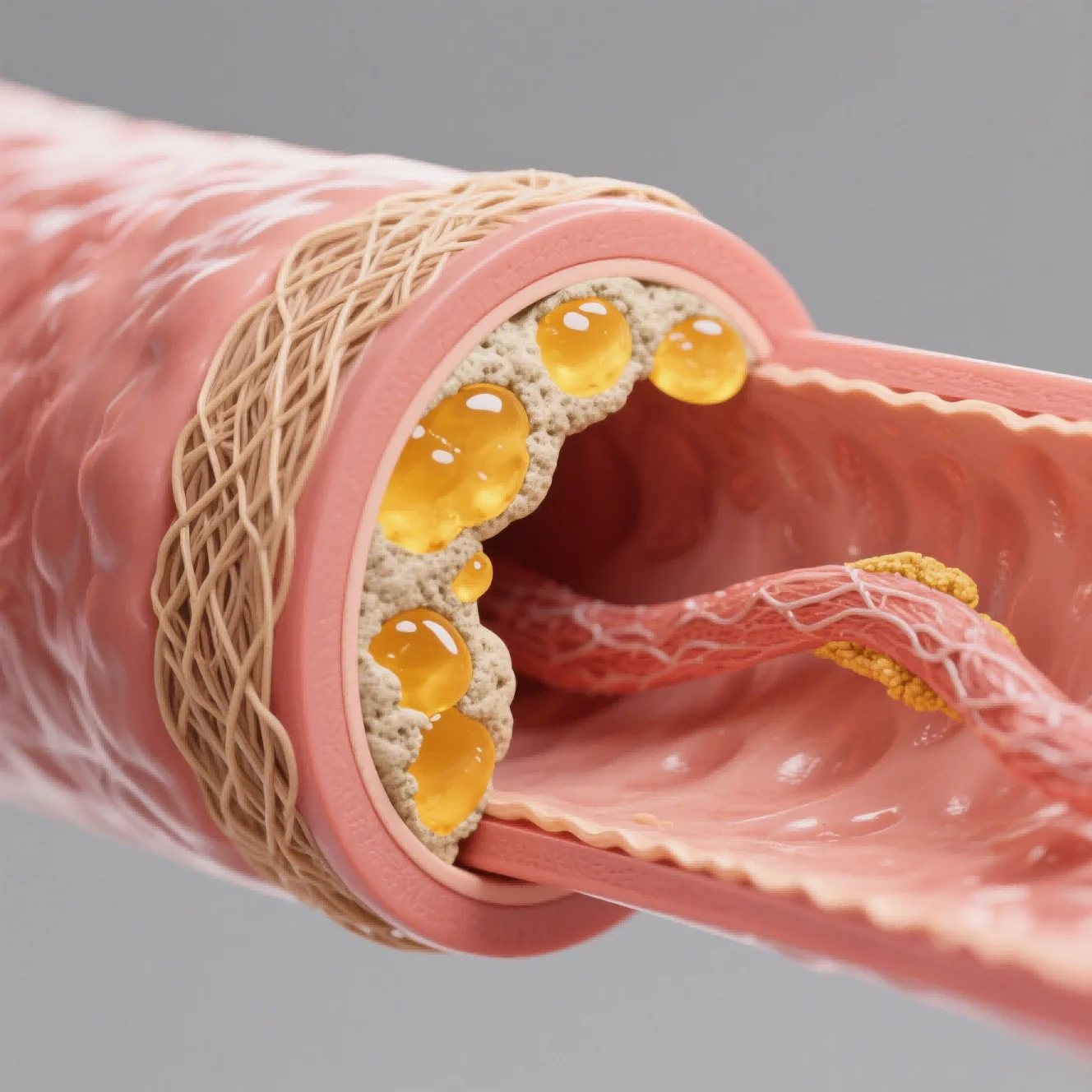
Liver-fibrosis research in 2026 is moving along five connected fronts: induction methods are becoming more mechanism-specific, studies follow a broader disease continuum, endpoint panels reach beyond a single stain, longitudinal observations are more common, and data delivery is easier to audit. These shifts are changing how animal models of liver fibrosis support antifibrotic candidate selection.
The shift responds to a familiar problem. Fibrosis is the outcome of interacting injury, inflammation, stellate-cell activation, matrix deposition, metabolism, and repair. A single induction method or terminal stain may reproduce only part of that process, so model value depends on which component the therapy is expected to change.
The planning question is specific: Does the candidate block an injury driver, reduce inflammation, alter metabolism, inhibit collagen deposition, or promote regression? The answer guides model choice, treatment start, duration, endpoints, and the reference therapy used to test responsiveness. Model selection preserves the causal link between intervention and the feature being measured.
Complexity carries a cost as well as a promise. Combined models can improve relevance but add variability and time; richer endpoints can clarify mechanism but increase multiplicity. Each design needs acceptance criteria and a decision rule that justify its added complexity. Feasibility work estimates the additional animals, samples, analysis time, and interpretation burden before the protocol is approved.
Trend 1: Mechanism-Matched Induction Models
Mechanism matching is becoming the entry point for model selection. Carbon tetrachloride creates reproducible toxic injury and fibrosis, while dietary systems foreground metabolic stress. An NASH-directed candidate and a compound acting downstream of repeated chemical injury begin with different induction logic. Animal models of liver fibrosis are more comparable after they are grouped by disease driver.
Combined induction is being used to represent multiple drivers, such as a high-fat diet plus a fibrogenic challenge. This may bring steatosis, inflammation, and fibrosis into one design, but the relative contribution of each driver must be understood. Otherwise, a response cannot be linked confidently to the intended mechanism.
Trend 2: Disease-Continuum Study Designs
Disease-continuum research follows obesity, insulin resistance, steatosis, steatohepatitis, fibrosis, and organ dysfunction as connected states. Treatment timing identifies whether the candidate prevents progression or acts on established disease. Added complexity earns its place only when that distinction changes the development decision.
Continuum designs require deliberate baselines and staging. Metabolic measures, liver injury markers, and histology can confirm the disease state before randomization or in matched sentinel animals. Without that confirmation, differences in induction timing may masquerade as therapeutic response. Sentinel findings also help place later tissue changes on a credible progression curve.
Trend 3: Multimodal Endpoint Panels
Layered endpoint sets separate liver injury from fibrogenic activity. AST and ALT describe injury; H&E maps architecture; Masson staining, α-SMA, and collagen measurements examine matrix deposition and stellate-cell activation. Metabolic, inflammatory, and molecular markers connect those tissue changes to mechanism. Across animal models of liver fibrosis, the hierarchy distinguishes injury, inflammation, matrix, and function.
Coordinating liver measurements across time is a practical platform test. Jennio Biotech brings chemically induced and diet-related fibrosis approaches together with biochemistry, H&E, Masson staining, α-SMA, collagen, imaging, and molecular analysis. Jennio Biotech can support that combination when collection timing and endpoint hierarchy are settled before the first cohort begins.
Trend 4: Longitudinal Monitoring Across Disease Progression
Longitudinal monitoring adds the missing time dimension. Repeated biochemistry, body weight, metabolic observations, or suitable imaging can reveal onset, plateau, regression, and rebound within the same animal. Historical biochemical and histological ranges help the team recognize an induction batch that drifts before treatment effects are interpreted.
Longitudinal designs still need terminal confirmation. At selected stages, pathology anchors imaging and circulating markers to tissue analysis. Calibration, consistent acquisition settings, and blinded review are necessary so that a time-course trend reflects biology rather than instrument or reader drift. Matched sampling times keep the circulating signal and tissue lesion biologically comparable.
Trend 5: Traceable and Integrated Study Delivery
Integrated delivery is the fifth shift. Induction, dosing, observation, bioanalysis, pathology, molecular assays, and statistics share one evidence map. Stable sample identifiers and timestamps preserve the route from exposure through tissue state to calculation, giving reviewers access to the chain behind each claim.
A liver-fibrosis delivery package keeps every layer connected: induction history, disease-stage evidence, exposure, enzyme data, tissue stains, molecular results, and statistical choices. That continuity lets a later reviewer retrace the biological argument without relying on a summary slide.
Cross-study metadata also helps reviewers detect model drift without treating historical cohorts, induction schedules, or endpoint panels as interchangeable. Versioned dictionaries preserve comparable endpoint definitions.
Independent review remains part of an integrated workflow. Pathology cross-checks, raw-data access, predefined exclusions, and electronic audit trails help protect the conclusion. The sponsor receives protocol history, induction and dosing records, specimen inventory, analytical outputs, representative and systematic images, and documented statistical decisions.
Regression studies may require a longer observation window than prevention studies using the same model. A second pathology reader can test whether borderline fibrosis grades remain stable under review.
By the end of 2026, the most informative fibrosis studies will not be identified by the largest endpoint panels. They will be the studies that place the candidate in the right disease stage, watch the response unfold over time, and preserve the link between exposure, tissue change, mechanism, and treatment decision.
