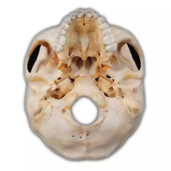
In the realm of anatomical education and medical practice, high-quality models are essential for effective learning and comprehension. The Digihuman 3D Printing Model–Skull Base represents a groundbreaking advancement in skull 3D printing technology. This anatomically precise model is crafted from high-precision digitized human body data, allowing for an accurate representation of the skull’s complex structures.
High-Precision Construction
The Digihuman Skull 3D printing utilizes full-color and multi-material 3D printers, employing environmentally friendly resin materials to create a 1:1 high simulation of physical anatomical models. This meticulous construction ensures that every detail of the skull base is represented accurately, from the texture of the uneven surfaces to the various anatomical features critical for understanding human anatomy. The model displays all holes, fissures, and protrusions in their true-to-life states, including sieve holes, rupture holes, spinous holes, and carotid tubes. Such precision is crucial for medical students and professionals who need to gain a thorough understanding of cranial anatomy.
Clear Visualization of Complex Structures
An essential advantage of the Digihuman Skull 3D printing model is its ability to vividly portray complex structures. Details such as the sphenoid pterygoid process, pear bone, inferior turbinate, and styloid process are clearly displayed, allowing users to study these intricate anatomical components effectively. Additionally, features like the foramen ovale, petrous part of the temporal bone, grooves, fissures, and impressions are distinguishable, facilitating deeper insights into skull-based anatomy. This high level of detail enhances the learning experience, making it invaluable for both educational and clinical applications.
In conclusion, the DIGIHUMAN 3D Printing Model-Skull Base presents a significant advancement in skull 3D printing technology. By providing an accurate, high-fidelity representation of the skull base, this model serves as an essential educational tool for students and professionals alike. Its detailed visualization capabilities not only foster a deeper understanding of cranial anatomy but also prepare future healthcare providers for real-world challenges in medical practice.


